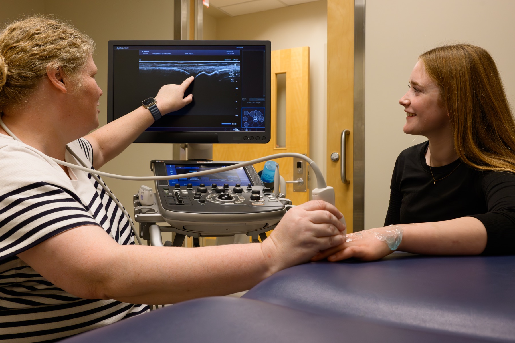
Juvenile idiopathic arthritis (JIA) affects bone development and joint health in children and adolescents, but its cause and progression are unpredictable. Our lab uses multimodal imaging to study changes in bone microarchitecture and the development of bone erosions, with the goal of detecting joint damage earlier and understanding how it progresses over time.
By comparing novel high-resolution imaging with conventional clinical imaging, we aim to improve the diagnosis, monitoring, and treatment of JIA while gaining new insights into how inflammatory arthritis affects the growing skeleton. This project aims to identify imaging biomarkers that can improve how joint health and bone development are monitored in children and adolescents with inflammatory arthritis.
Project Details
Related selected publication: Carpal bone segmentation for HR-pQCT imaging.
Explore trainee opportunities or contact us about this project.