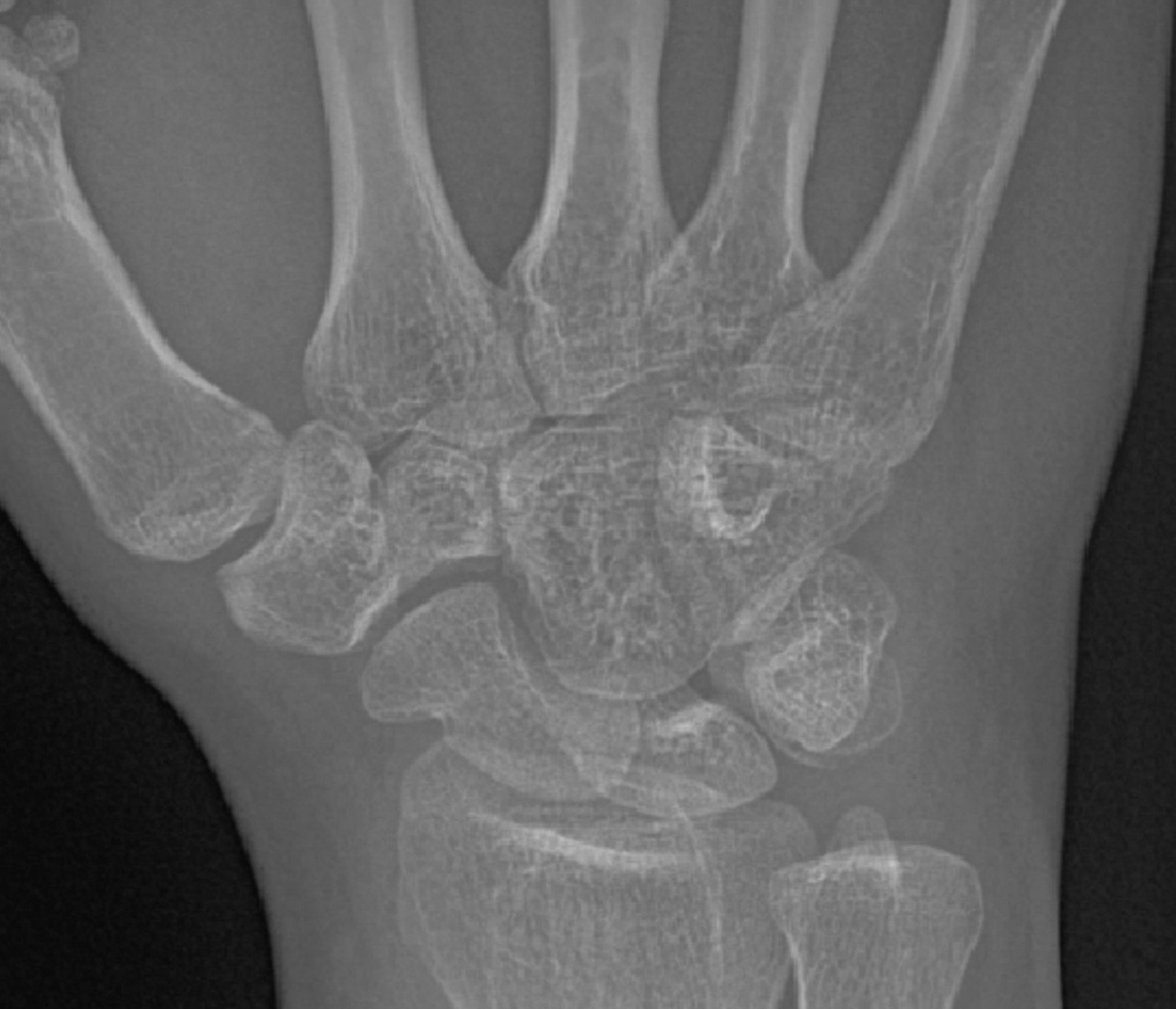
In vivo assessment of pediatric fracture healing
How does bone repair itself during growth? We use high-resolution imaging to follow fracture healing and understand differences in recovery.

Our work connects clinical imaging and preclinical bone mechanobiology to understand how growing bones develop, adapt, and recover.
How does bone repair itself during growth? We use high-resolution imaging to follow fracture healing and understand differences in recovery.
This project uses advanced musculoskeletal imaging and computational modelling to study why children with obesity can have elevated fracture risk despite higher bone mineral density.
This project uses multimodal imaging to study bone microarchitecture and erosions in juvenile idiopathic arthritis, with the goal of improving diagnosis, monitoring, and treatment.
We combine longitudinal pediatric cohorts, multimodal imaging, and computational analysis to understand bone growth, joint health, and recovery in children and adolescents.
How do mechanical forces guide bone growth? We study the response from cellular changes to whole-bone development.
Using experimental models, high-resolution microCT, controlled loading, and image-based biomechanics, we study how growing bones sense and respond to their mechanical environment.
Our clinical and preclinical imaging platforms, mechanical testing systems, and 3D printing resources support research from tissue-level structure to whole-bone health.
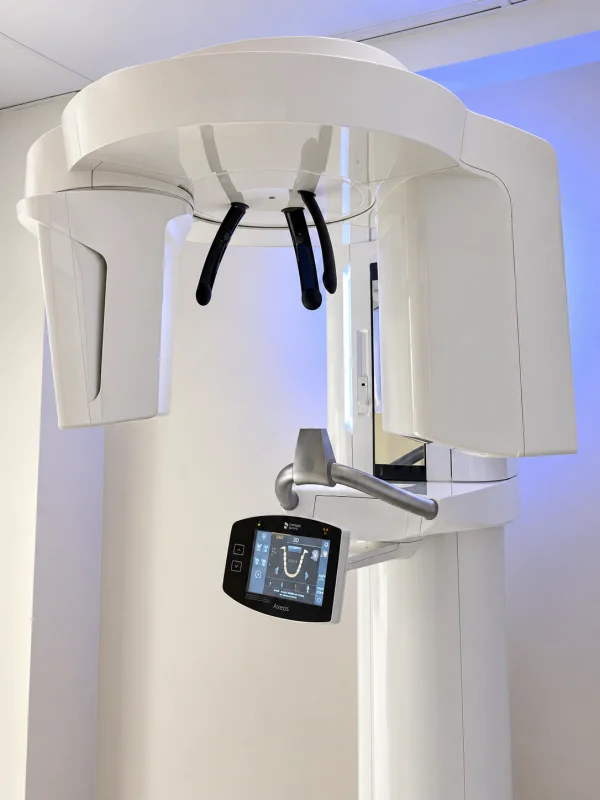
Axeos · Dentsply Sirona
In the lab
Three-dimensional imaging of mineralized structures to study bone shape and spatial anatomy.
Axeos combines two-dimensional X-ray imaging with three-dimensional cone beam CT. Selectable scan volumes and high-definition imaging allow mineralized structures to be visualized in detail and examined from multiple angles. These capabilities provide a platform for studying bone shape and spatial anatomy in our musculoskeletal research.
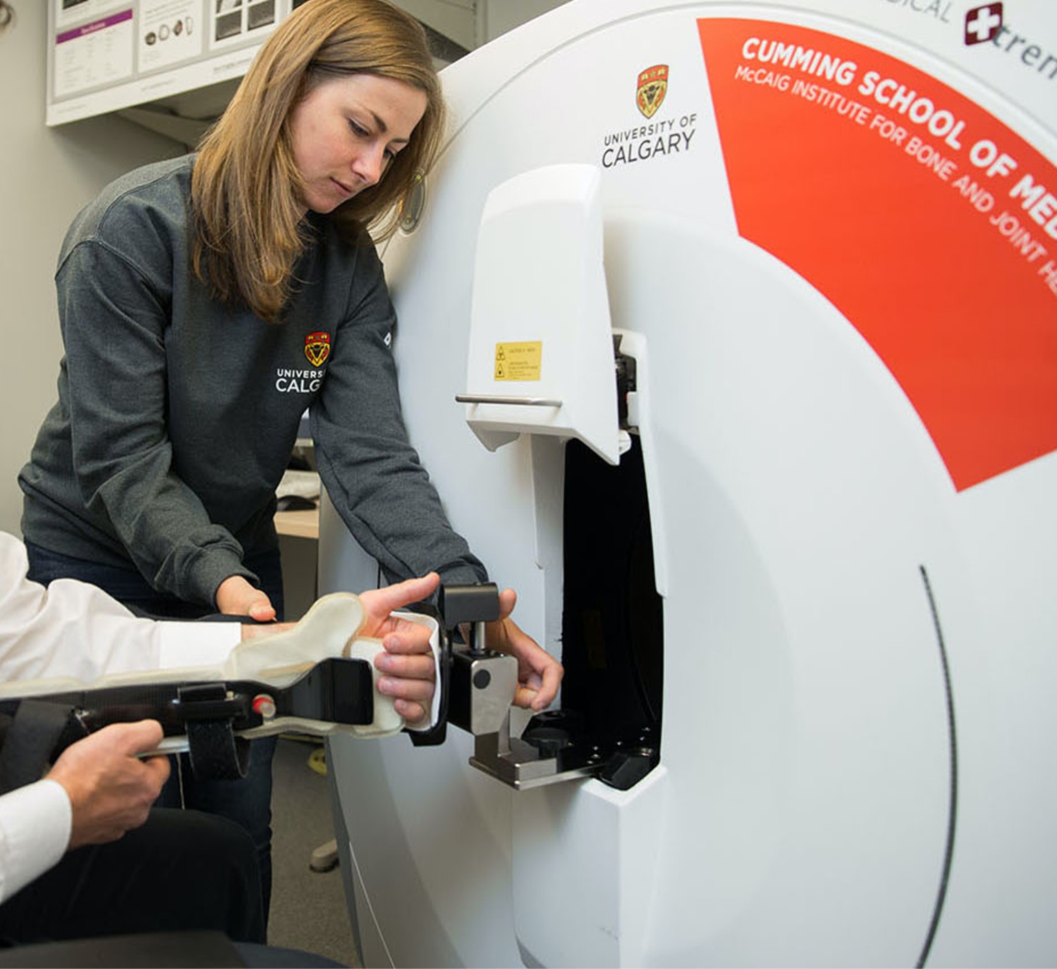
XtremeCT II · Scanco Medical
Access through the Centre for Mobility and Joint Health
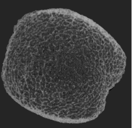
Detailed measurements of bone density and cortical and trabecular microarchitecture in peripheral bones.
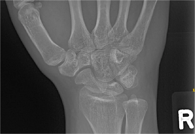
High-resolution peripheral quantitative CT produces detailed three-dimensional images of peripheral bone, capturing bone density and cortical and trabecular microarchitecture in a single scan. These measurements support research into skeletal development and changes in bone structure that can be difficult to see with conventional imaging. Access is provided through the Centre for Mobility and Joint Health.
Partner facility access
Complementary radiography, CT, DXA, and ultrasound for joint health and body composition research.
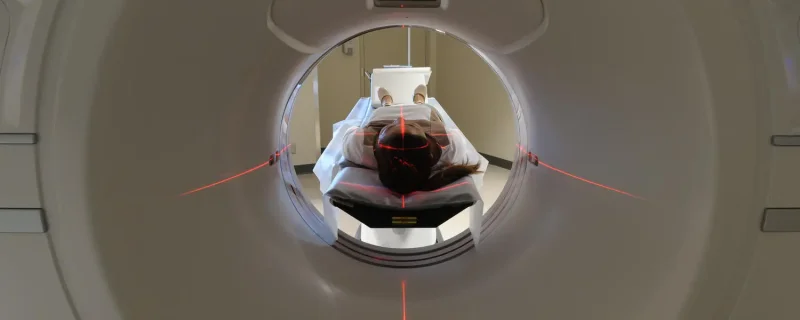
The Centre for Mobility and Joint Health provides access to complementary imaging platforms, including radiography, CT, DXA, and ultrasound. Combining these approaches can connect detailed bone structure with broader measurements of joint health and body composition. See the facility's equipment page for system details and current availability.
vivaCT80 · Scanco Medical
Access through the microCT Core
Repeated three-dimensional imaging to follow bone growth and adaptation in small-animal models.
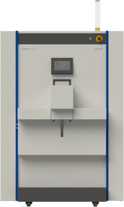
The vivaCT80 generates three-dimensional images of mineralized tissue in small-animal models. Repeated imaging supports longitudinal studies of bone growth, adaptation, and responses to experimental conditions, allowing structural changes to be followed over time. Access is provided through the microCT Core.
µCT50/µCT100 · Scanco Medical
Access through the microCT Core
High-resolution imaging of isolated specimens for quantitative analysis of cortical and trabecular architecture.
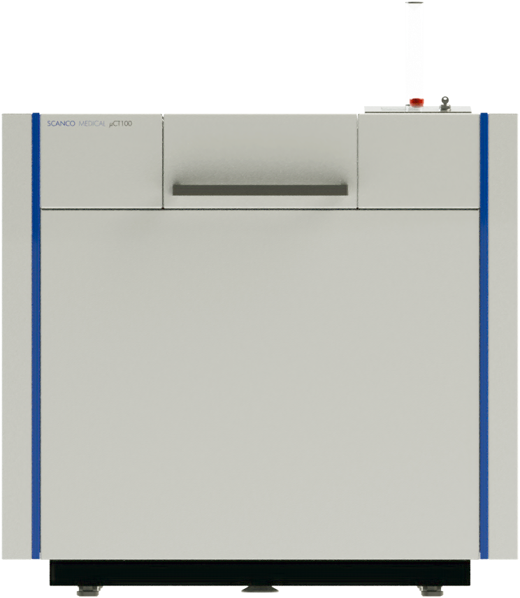
The µCT50 and µCT100 systems provide high-resolution, non-destructive three-dimensional imaging of isolated bone and other specimens. Their detailed images support quantitative analysis of cortical and trabecular architecture, helping connect tissue structure with biological and mechanical measurements. Access is provided through the microCT Core.
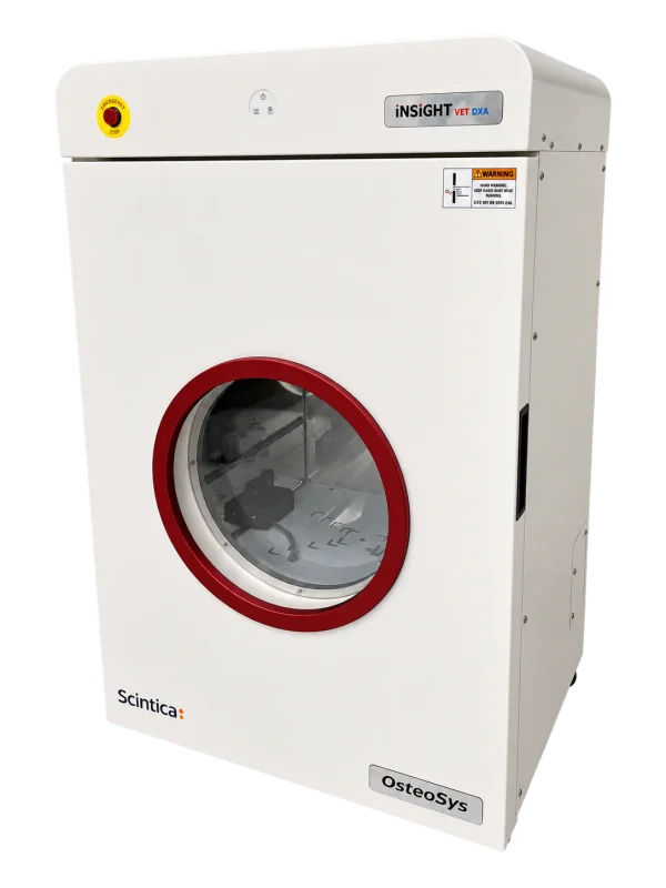
iNSiGHT · Scintica
In the lab
Bone mineral density, bone mineral content, and body composition measurements in small animals.
iNSiGHT uses dual-energy X-ray absorptiometry to measure bone mineral density, bone mineral content, and body composition in small animals. Estimates of fat and lean mass complement bone measurements, supporting studies of skeletal growth and metabolic influences on bone health. The system can be used to track these measurements across a longitudinal study.
BioDent · ActiveLife Scientific
In the lab
Local indentation measurements to examine how bone tissue responds to mechanical loading.
BioDent uses an outer reference probe and an inner test probe to measure local indentation of a bone surface. Repeated loading cycles record tissue deformation relative to the reference point, providing information about bone's response to mechanical loading. These tissue-level measurements complement imaging of bone geometry and microarchitecture.
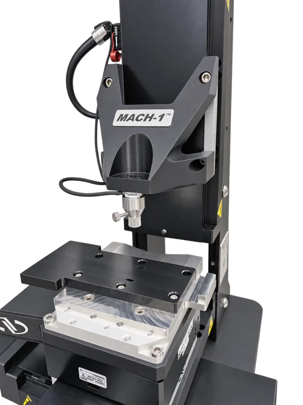
Mach-1 V500c · Biomomentum
In the lab
Controlled mechanical testing to connect tissue structure with stiffness and material behaviour.
Mach-1 measures how tissues and biomaterials respond to controlled mechanical loading. With suitable fixtures and protocols, compression and indentation tests can assess stiffness and time-dependent deformation through force and displacement measurements. These tests help connect tissue structure with mechanical function and evaluate how materials behave under load.
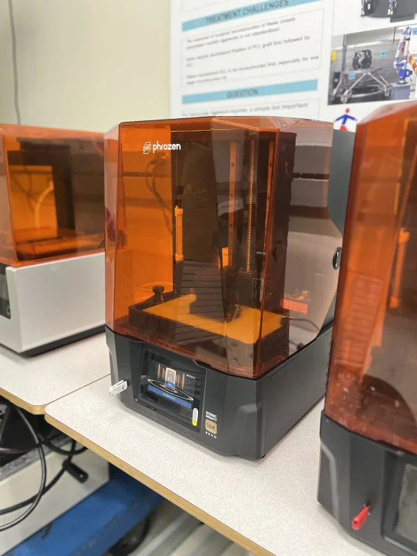
Sonic Mighty Revo 16K · Phrozen
In the lab
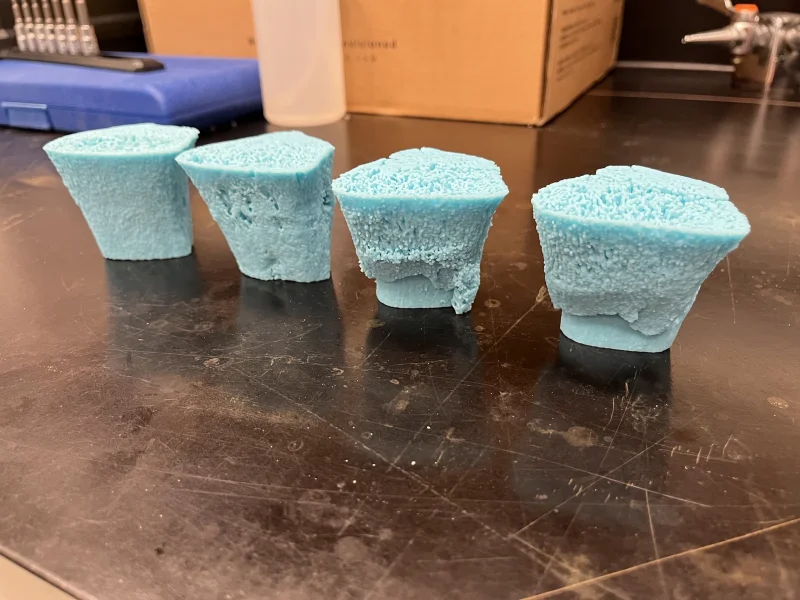
Detailed physical bone models for research communication, hands-on teaching, and prototyping.
The Sonic Mighty Revo 16K is an LCD-based resin 3D printer designed to produce detailed physical models. Its high-resolution printing capabilities support the translation of imaging-derived bone models into tangible objects with fine structural features. Printed models can support research communication, hands-on teaching, and prototype development, helping make complex skeletal anatomy easier to explore.
Interested in collaborating or using our lab's resources? Contact us at pediatricmsk@ucalgary.ca to discuss opportunities.